
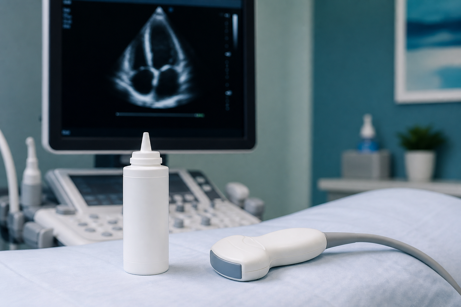
An echocardiogram is an ultrasound of the heart. It uses sound waves to show chamber size, pumping strength, valve motion, and how blood moves through the heart. It does not use ionizing radiation the way a CT scan does. Cardiologists order it when they need structure and function details that a stethoscope and ECG alone cannot provide.
People often search after a new murmur, unexplained shortness of breath, swelling, abnormal ECG, or a family history of heart muscle disease. Others want an echocardiogram near me or echocardiogram in Plano because a primary-care clinician already suggested imaging. This guide explains what the test shows, how long it takes, what it does not diagnose, and how results guide next steps.
If your question is about rhythm over a full day rather than heart structure, start with what a Holter monitor is. For broader heart services, see cardiac services and Plano location details.
What an echocardiogram images
A standard transthoracic echocardiogram (TTE) looks through the chest wall with a probe and gel. The report usually comments on:
- Left and right ventricle size and wall motion
- Ejection fraction or other measures of pumping strength
- Valve opening, closing, and leak or narrowing
- Estimated pressures and filling patterns when measurable
- Pericardial fluid around the heart if present
An echocardiogram images valves, chambers, and pump function. It does not reliably map most coronary artery blockages the way a stress test pathway or coronary CT/angiography might. That distinction prevents false expectations.
Sonographers also capture multiple views so the reading cardiologist can cross-check findings. One limited window is rarely enough for a complete study. That is why body habitus, prior surgery, and lung disease can make some images harder and occasionally lead to a recommendation for a different echo type.
Why cardiologists order one
Common reasons include a murmur, heart failure symptoms, cardiomyopathy screening, valve follow-up, unexplained edema, or abnormal ECG findings that need structural context. After chemotherapy or other treatments that can affect the heart, serial echoes may track function over time.
Ordering an echo is not an automatic sign of a crisis. It is often a careful next step when history and exam leave an open question about structure or pumping strength.
Primary care clinicians also order echocardiograms when they hear a new murmur or see swelling that needs a cardiology-level look at pump function. The goal is the same: answer a specific structural question before escalating to more invasive testing.
What to expect during the test
You lie on an exam table, usually on your left side for parts of the study. A sonographer places gel on the chest and moves the probe to different windows between the ribs. You may hear Doppler sounds as blood flow is measured. The room is dim so the screen is easier to see.
Most transthoracic studies take about 30 to 60 minutes, depending on body habitus, study complexity, and whether contrast is used. You can usually resume normal activity afterward. There is no recovery downtime like sedation-based procedures.
Prep tips
- Wear a two-piece outfit so the chest is easy to access
- Bring a medication list and prior heart reports if you have them
- Tell staff about recent surgeries, implants, or breathing limits that affect positioning
- Ask whether fasting is required; many TTEs do not need it
Echocardiogram vs Holter vs stress test
- Echocardiogram: structure and pump function with ultrasound
- Holter / patch monitor: rhythm over hours to weeks
- Stress test: how the heart responds to exercise or medication stress, sometimes with imaging
These tools answer different questions. Many patients need more than one over time. Your clinician chooses based on symptoms, risk, and what the first tests already showed.
If flutters or skipped beats are the main complaint, rhythm monitoring may come first or in parallel. If the concern is valve disease or reduced pumping strength, echo usually leads. Ask which question today’s order is trying to answer so you know what “normal” or “abnormal” will mean for next steps.
What results can mean
Results may be reassuringly normal, show mild age-related changes, or identify valve disease, reduced pumping strength, thickened walls, or fluid around the heart. “Abnormal” covers a wide range. Mild findings may only need monitoring. Significant findings can lead to medication changes, blood pressure optimization, further testing, or specialist follow-up.
Ask three practical questions at results review:
- Which finding matters most for my symptoms?
- Does this change treatment now or only watching?
- Do I need another test, such as stress imaging or longer rhythm monitoring?
Special types you might hear about
A stress echocardiogram combines ultrasound with exercise or medication stress. A transesophageal echocardiogram (TEE) places a probe in the esophagus for clearer views of some structures when TTE windows are limited. Those studies have different prep and recovery rules. Your order will say which type you are scheduled for.
Getting an echocardiogram in Plano
Prime Heart and Vascular evaluates patients who need heart imaging as part of preventive and diagnostic cardiology. If you were told to get an echocardiogram near you, bring the reason for the order, prior records, and your symptom timeline. You can schedule an appointment to discuss whether echo, Holter monitoring, or another test fits first.
Questions to ask before you schedule
Ask whether the order is transthoracic, stress echo, or TEE. Ask if prior outside images should be brought on a disc or portal download. Ask whether you should take usual morning medicines. Clear answers prevent a wasted trip and a repeat study.
If anxiety about the test is high, say so. Knowing the probe stays on the chest wall for a standard TTE helps many people relax. TEE is different and deserves a separate explanation from the ordering clinician.
After the echocardiogram: making results actionable
A report sitting unread in a portal does not help. Schedule a results conversation when the study is ordered, or ask who will call you. Bring a short symptom list so the interpreter can connect findings to what you feel climbing stairs or lying flat.
If the echo is normal but symptoms continue, that is still useful information. It may shift attention toward rhythm monitoring, lung causes, deconditioning, or another pathway. Normal structure does not mean the visit was wasted.
Keep copies of key reports. Outside hospitals and new clinics often need prior ejection fraction and valve details to avoid repeating identical studies too soon.
American Heart Association educational materials continue to emphasize imaging when history and exam leave important structural questions open. Your own symptoms and risk still decide timing more than any generic checklist.
Bring questions in writing so the results visit covers treatment changes, monitoring intervals, and whether family members need screening for inherited cardiomyopathy when relevant.
Limits of echocardiogram and when other tests follow
An echocardiogram is powerful for structure and pump function, but it is not a complete heart evaluation by itself. Chest pain that sounds like coronary disease may still need stress testing or anatomic coronary imaging. Palpitations may need a Holter or event monitor even when the echo looks normal. Shortness of breath can involve lungs, anemia, deconditioning, or valve disease, so clinicians often combine history with more than one test.
Knowing those limits helps you avoid false reassurance or false alarm. Ask what question the echo answered and what question remains open. That conversation is how imaging becomes a plan instead of a PDF in a portal.
Putting the pieces together
An echocardiogram is ultrasound of the heart used to assess chambers, valves, and pumping strength without ionizing radiation. It is a structure test, not a full map of coronary blockages. Prep is usually simple, the study often lasts under an hour, and results should turn into a clear next step with your cardiology team.
Schedule an appointment with Prime Heart and Vascular to discuss whether an echocardiogram or another heart test is the right next step.
Echocardiogram questions
An echocardiogram is an ultrasound picture of your heart. A probe on the chest uses sound waves to show how the chambers move, how strong the pump is, and how the valves work. It does not use X-ray radiation. Clinicians order it when they need structural detail beyond an exam and ECG. Results help decide whether to watch, treat, or test further.
A typical transthoracic echocardiogram takes about 30 to 60 minutes. Body size, image quality, and whether extra views or contrast are needed can change the timing. You usually go home and resume normal activity right after. Ask the scheduling team if your order is a stress echo or TEE, because those visits take longer and have different prep.
A standard echo does not map most coronary blockages the way angiography or some CT pathways can. It shows chamber function, valves, and pumping strength. Wall-motion clues can sometimes raise concern, but they are not a full artery map. If blocked arteries are the main question, your clinician may order a stress test or other coronary evaluation instead of or after an echo.
No. An ECG records electrical signals and rhythm at a moment in time. An echocardiogram creates ultrasound images of heart structure and motion. Many patients get both because rhythm and structure answer different questions. An abnormal ECG is one common reason an echo is ordered, but a normal ECG does not always mean an echo is unnecessary when symptoms point to structural problems.
For most transthoracic echoes, wear clothes that open easily at the chest and bring your medication list. Fasting is often not required, but follow the instructions on your order. Tell staff about implants, recent surgery, or trouble lying on one side. Stress echo and TEE have stricter prep rules, including possible fasting and medication holds, so confirm before the visit.
A sonographer acquires images, then a cardiologist interprets the study. Timing varies by clinic workflow. Some results return within a few days; complex studies may take longer. Do not treat a portal preview as final advice without clinician context. Ask who will call you and whether a follow-up visit is planned to explain findings.
Yes. Local cardiology practices, including Prime Heart and Vascular, arrange heart ultrasound when clinically indicated. Bring the reason for the order and prior records. If you searched echocardiogram near me after a primary-care suggestion, schedule a visit so the team can confirm the right test type and timing. Urgent symptoms such as severe chest pain still belong in emergency care first.
Echo looks at structure and pump function with ultrasound. Holter monitoring records rhythm over a longer outdoor period. Fluttering, skips, or racing spells often need rhythm capture. Murmurs, pump concerns, or valve questions often need echo. Your history decides which comes first and whether both are useful.